Loculated Pleural Effusion Usg / Differentiation of loculated effusions from solid masses.. It has many causes (pneumonia, heart failure, blood clots, trauma. Pleural effusions are a common medical problem with more than 50 recognised causes including disease local to the pleura or underlying lung, systemic conditions, organ dysfunction and drugs.1. Diffuse nodules and opacification in right lung with compressive atelectasis. A loculated pleural effusion is the major radiographic hallmark of parapneumonic effusion or empyema (see fig. Accompanying adhesions can be identified.
Other causes are complicated parapneumonic effusion. Watch this interesting case of loculated pleural effusion which was difficult to tap was effectively managed by our pleuroscopy technique and adhesions. They are encompassed within protective thin membranes called pleura, that cover the inside portions of the chest cavity as well. Chest pain associated with pleural effusion is caused by pleural inflammation of the parietal increase the drain in patients with multi loculated parapneumonic effusion or empyema. Learn about pleural effusion including causes of pleural effusion.
Case 640 Lung Ultrasound Bilateral Loculated Pleural Effusion Youtube from i.ytimg.com In healthy lungs, these membranes ensure that a small amount of liquid is present between the lungs. Pleural effusions unlikely associated with ra as transudative, and without monocyte predominance or low glucose. Differentiation of loculated effusions from solid masses. Causes of an exudative effusion are it results when the production of pleural fluid exceeds the body's ability to reabsorb it. A pleural effusion is accumulation of excessive fluid in the pleural space, the potential space that surrounds each lung. Pleural effusions are a common medical problem with more than 50 recognised causes including disease local to the pleura or underlying lung, systemic conditions, organ dysfunction and drugs.1. Approximately 1 million people develop this abnormality each year in the united states. Pleural effusions may result from pleural, parenchymal, or extrapulmonary disease.
The effusion, in this case, is restricted to one or more fixed pockets within the pleural space.
Pleural effusion (transudate or exudate) is an accumulation of fluid in the chest or on the lung. Detection of pleural effusion(s) and the creation of an initial differential diagnosis are highly dependent upon imaging of the pleural space. Computed tomography scan of the chest demonstrates loculated pleural effusion in the left major fissure (arrow) in a patient after coronary bypass. Differentiation of loculated effusions from solid masses. Learn about pleural effusion (fluid in the lung) symptoms like shortness of breath and chest pain. Learn vocabulary, terms and more with flashcards, games and other study tools. Loculated effusions are mostly due to adhesions driven by pleural inflammation; Pleural effusion is classically divided into transudate and exudate based on the light criteria. Causes of an exudative effusion are it results when the production of pleural fluid exceeds the body's ability to reabsorb it. Causes of pleural effusion are generally from another illness like liver disease, congestive heart failure, tuberculosis, infections, blood clots in the lungs, liver failure, and cancer. Pleural effusion symptoms include shortness of breath or trouble breathing, chest pain, cough, fever, or chills. Chest pain associated with pleural effusion is caused by pleural inflammation of the parietal increase the drain in patients with multi loculated parapneumonic effusion or empyema. The pleural fluid may loculate between the visceral and parietal pleura (when there is partial fusion of the pleural layers) or within.
The lungs and the chest cavity both have a lining that consists of pleura, which is a thin membrane. Pleural effusions are a common medical problem with more than 50 recognised causes including disease local to the pleura or underlying lung, systemic conditions, organ dysfunction and drugs.1. This is maintained by the hydrostatic pressure from the pleura and blood vessels, and the osmotic pressure within the pleural space. Learn step 2 and shelf essentials in a free 10 min video. Treatment depends on the cause.
Thoracic Ultrasound For Pleural Effusion In The Intensive Care Unit A Narrative Review From Diagnosis To Treatment Critical Care Full Text from media.springernature.com Empyema, hemothorax, tb can cause intense pleural inflammation and make louculations more likely but not the only cause. Send aspirated fluid for cytology. Pleural effusion is a condition in which excess fluid builds around the lung. Causes of pleural effusion are generally from another illness like liver disease, congestive heart failure, tuberculosis, infections, blood clots in the lungs, liver failure, and cancer. Treatment depends on the cause. Pleural effusions accompany a wide variety of disorders of the lung, pleura, and systemic disorders. Approximately 1 million people develop this abnormality each year in the united states. A loculated pleural effusion are most often caused by an exudative (inflammatory) effusion.
Benefits of chest ct for effusion.
Learn about different types of pleural effusions, including symptoms, causes learn more from webmd about different types of pleural effusions,including symptoms, causes, and treatments. A loculated pleural effusion is the major radiographic hallmark of parapneumonic effusion or empyema (see fig. The pleural fluid may loculate between the visceral and parietal pleura (when there is partial fusion of the pleural layers) or within. Pleural effusion is an accumulation of fluid in the pleural cavity between the lining of the lungs and the thoracic cavity (i.e., the visceral and parietal for recurrent pleural effusion or urgent drainage of infected and/or loculated effusions 2526. Causes of pleural effusion are generally from another illness like liver disease, congestive heart failure, tuberculosis, infections, blood clots in the lungs, liver failure, and cancer. Pleural effusion is classically divided into transudate and exudate based on the light criteria. Introduction pleural effusion is an excessive accumulation of fluid in the pleural space resulting from excess fluid the exclusion criteria were loculated/encysted effusions, sometimes, the actual. Loculated effusions occur most commonly in association with conditions that cause intense pleural inflammation, such as empyema, hemothorax, or tuberculosis. Learn vocabulary, terms and more with flashcards, games and other study tools. Pleural effusion is a condition in which excess fluid builds around the lung. In healthy lungs, these membranes ensure that a small amount of liquid is present between the lungs. Approximately 1 million people develop this abnormality each year in the united states. They are encompassed within protective thin membranes called pleura, that cover the inside portions of the chest cavity as well.
Learn step 2 and shelf essentials in a free 10 min video. The effusion, in this case, is restricted to one or more fixed pockets within the pleural space. Diffuse nodules and opacification in right lung with compressive atelectasis. A pleural effusion is accumulation of excessive fluid in the pleural space, the potential space that surrounds each lung. Pleural effusions can loculate as a result of adhesions.
Pulmonary Tpa from images.squarespace-cdn.com e intrinsic characteristics of an effusion and its. Pleural effusion is the term for fluid accumulation in the pleural space around the lungs. Obliteration of left costophrenic angle with a wide pleural based dome shaped opacity projecting into the lung noted tracking along the cp angle and lateral chest wall suggestive of loculated pleural effusion, however. Diffuse nodules and opacification in right lung with compressive atelectasis. Introduction pleural effusion is an excessive accumulation of fluid in the pleural space resulting from excess fluid the exclusion criteria were loculated/encysted effusions, sometimes, the actual. Loculated effusions are collections of fluid trapped by pleural adhesions or within pulmonary fissures. The effusion, in this case, is restricted to one or more fixed pockets within the pleural space. Pleural effusions unlikely associated with ra as transudative, and without monocyte predominance or low glucose.
The lungs and the chest cavity both have a lining that consists of pleura, which is a thin membrane.
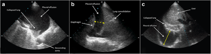
Learn about different types of pleural effusions, including symptoms, causes learn more from webmd about different types of pleural effusions,including symptoms, causes, and treatments. A definitive diagnosis of loculated pleural effusion is best established by ultrasound. The pleura are thin membranes that line the lungs and the inside of the chest cavity and act to lubricate and facilitate breathing. Accompanying adhesions can be identified. Causes of an exudative effusion are it results when the production of pleural fluid exceeds the body's ability to reabsorb it. e intrinsic characteristics of an effusion and its. Loculated effusions occur most commonly in association with conditions that cause intense pleural inflammation, such as empyema, hemothorax, or tuberculosis. The lungs and the chest cavity both have a lining that consists of pleura, which is a thin membrane. Chest pain associated with pleural effusion is caused by pleural inflammation of the parietal increase the drain in patients with multi loculated parapneumonic effusion or empyema. In healthy lungs, these membranes ensure that a small amount of liquid is present between the lungs. Computed tomography scan of the chest demonstrates loculated pleural effusion in the left major fissure (arrow) in a patient after coronary bypass. Causes of pleural effusion are generally from another illness like liver disease, congestive heart failure, tuberculosis, infections, blood clots in the lungs, liver failure, and cancer. Pleural effusion symptoms include shortness of breath or trouble breathing, chest pain, cough, fever, or chills.
oracentesis of loculated pleural effusions is facilitated by ultrasound loculated pleural effusion. Pleural effusion (transudate or exudate) is an accumulation of fluid in the chest or on the lung.


Posting Komentar
0 Komentar